Micro procedures.
Natural reinforcement.
Breakthrough micro-instrumentation for high-precision, ab interno surgery — and natural allogeneic tissue that reinforces the eye's own structure. Better together.
Breakthrough micro-instrumentation.
Micro-scale instruments engineered to reach the angle through a tiny opening — enabling canaloplasty, excisional goniotomy, and cyclodialysis, and delivering tissue with single-pass precision.
AlloSert™ Uveo
Microinterventional cyclodialysis and scleral-graft delivery platform.
Breakthrough bio-tissue products.
Structural by design, made to reinforce. A single tissue platform, shaped for the anatomy it supports — from the peri-canal wall to endoscleral reinforcement.
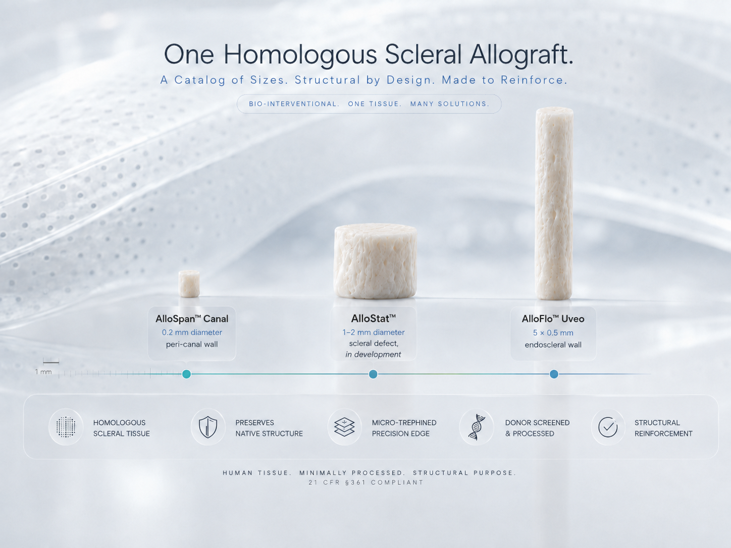
Natural allogeneic bio-reinforcement.
Instead of permanent synthetic hardware, the surgical site is reinforced with the eye's own kind of tissue — minimally manipulated human scleral allograft that provides natural, structural reinforcement of native sclera.
- The same collagenous tissue as the host sclera
- Matched biomechanics — no rigidity mismatch with native sclera
- Porous, conforming scleral matrix
- No permanent synthetic material left in the eye
- Human tissue under §361 — exempt from device premarket approval
- Foreign, engineered material
- Stiffness mismatch with the soft eye
- Permanent, rigid implant
- A device — cleared or approved via 510(k) or PMA
Because homologous sclera shares native tissue's mechanics, it avoids the stiffness mismatch that makes rigid synthetic implants a recognized trigger of the foreign-body response. It isn't a device by nature — which is why it isn't one by regulation: homologous human tissue is exempt from device premarket approval under §361.
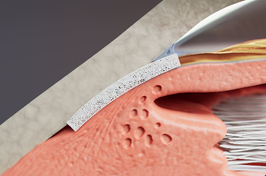
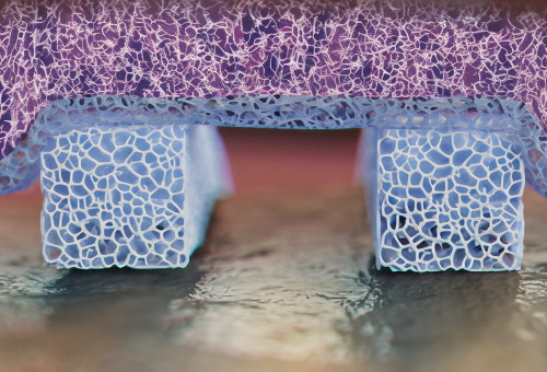

AlloFlo™ Uveo
Gentle, minimally processed human scleral tissue that reinforces the eye's own endoscleral wall at the surgical site.
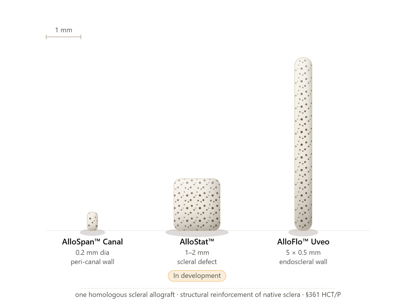
- AlloSpan™ Canal0.2 mm dia · peri-canal wall
- AlloStat™ In development1–2 mm · scleral defect
- AlloFlo™ Uveo5 × 0.5 mm · endoscleral wall
Human tissue, handled to standard.
Recovered from consented donors and processed by an accredited eye bank.
Enabling breakthrough procedures for the modern surgeon.
The instrument performs the procedure and places the tissue exactly where it's needed; the tissue reinforces the eye's own wall. Together, they let the modern surgeon complete an ab interno, tissue-sparing procedure with natural reinforcement — and leave no permanent synthetic device behind.
Access
A single micro-instrument reaches the angle through a tiny ab interno opening.
Perform the procedure
The surgeon performs canaloplasty, goniotomy, or cyclodialysis using the eye's natural anatomy; the tissue provides structural reinforcement only.
Reinforce
Natural scleral tissue is delivered and reinforces the surgical site — no permanent synthetic device left behind.
Instrument and tissue, engineered as one workflow. Better together.
Procedures & reinforcement.
A single-pass platform across the angle-based procedures you already perform — with the option of natural scleral reinforcement.
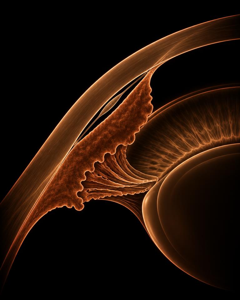
Cyclodialysis
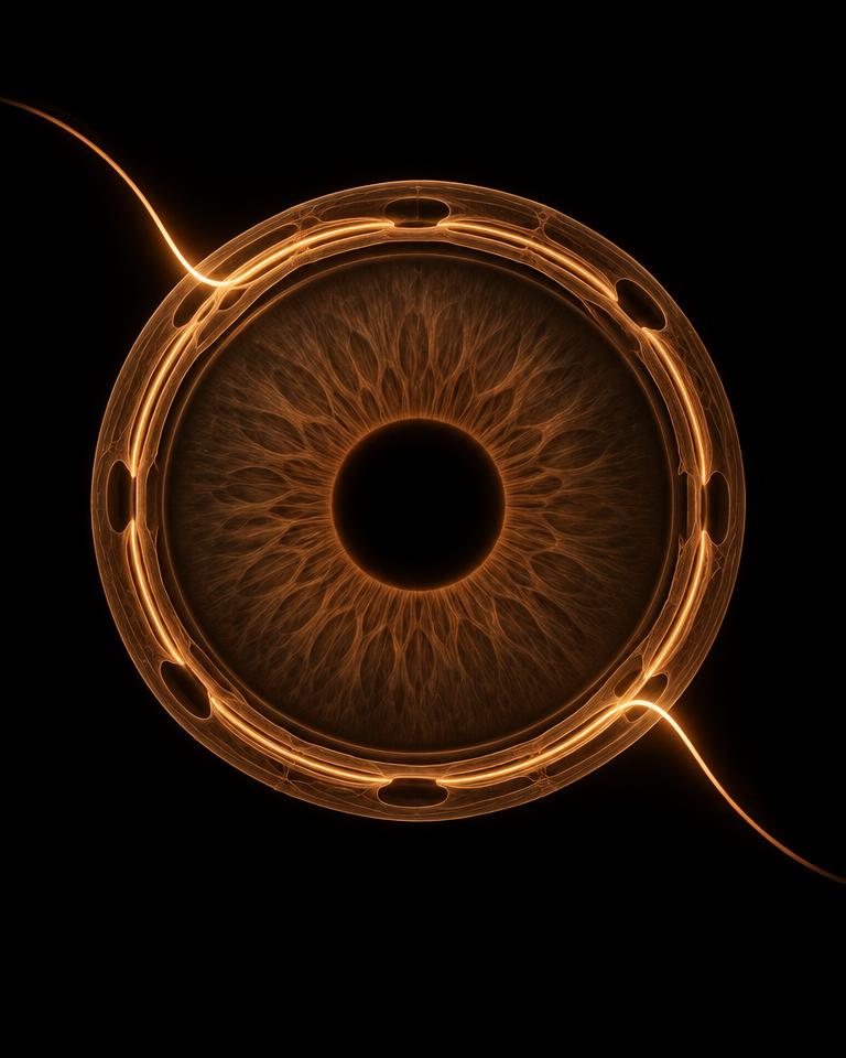
Canaloplasty
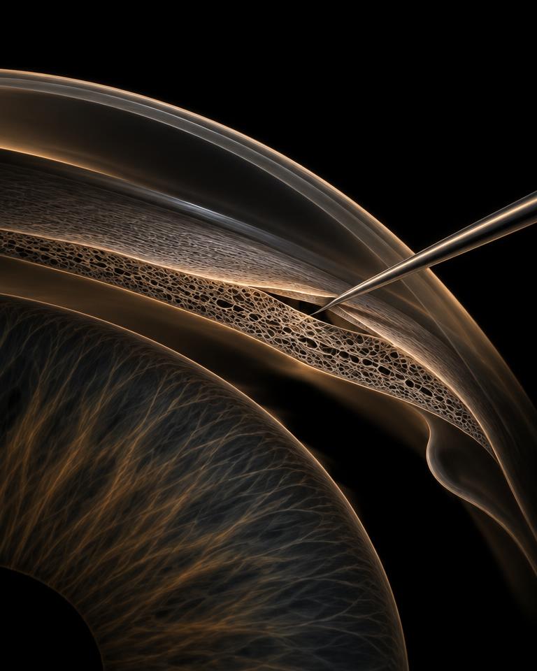
Goniotomy

Optic disc pit surgery
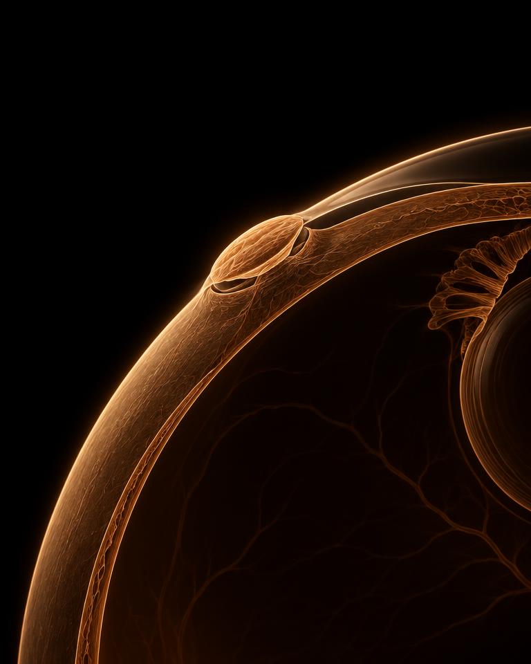
Sclerostomy closure
The allograft provides homologous structural reinforcement of native sclera. Optic disc pit and sclerostomy applications, and AlloStat™, are in development and not available for commercial sale.
Clinical literature.
Peer-reviewed publications on the cyclodialysis and canaloplasty procedures, including the CREST study — one of the largest prospective cohorts in interventional glaucoma.
Outcomes below are reported for the surgical procedures as published; findings and terminology are the authors' own and are not claims for any tissue or instrument.
Reiss et al. PLoS One. 2026;21:e0351552.
Ianchulev T, Weinreb RN, Calvo E, et al. Clin Ophthalmol. 2024;18:3605–3614.
Chaya CJ, Herndon LW, Lince J, et al. J Clin Med. 2024;13(16):4593.
Calvo E, De Francesco T, Vera L, Tyson F, Weinreb RN. Ophthalmol Sci. 2025;5(4):100727.
Stamper R, Huang A, Toris C, et al. Clin Ophthalmol. 2025;19:2859–2870.
De Francesco T, Ianchulev T, Rhee DJ, et al. Clin Ophthalmol. 2024;18:1789–1795.
Barber K, Flowers B, Patterson M, Vera L, Ahmed IIK. Ther Adv Ophthalmol. 2025;17.
Ianchulev T, et al. Br J Ophthalmol. 2024;108:217–222.
These publications evaluate the surgical procedure. The scleral allograft's role within the procedure is structural reinforcement of native sclera. Full presentation and CREST materials are available to healthcare professionals on request.
Iantrek in the field.
Independent coverage of micro-interventional glaucoma surgery and angle-based procedures.
The Evolution of Canaloplasty
Expanding options in the suprachoroidal space
Should surgeons target both outflow pathways?
External coverage published by third parties. Iantrek does not control third-party content.
See the procedure.
A single-pass, ab interno approach — with optional natural scleral reinforcement.
- Ab interno accessReach the angle through a single micro-incision.
- Single-pass efficiencyCanaloplasty with excisional goniotomy in one instrument pass with C-Rex.
- Fits the cataract workflowIntegrates with standard phacoemulsification.
- Optional reinforcementDeliver natural scleral tissue to reinforce the surgical site.
Published in the peer-reviewed literature.
The clinical experience of surgeons working with this platform is reported independently, in peer-reviewed journals — the compliant, durable record of the field's evidence.
The following are independent, peer-reviewed publications, listed for scientific and educational reference. Their findings, methods, and terminology are the authors' own. Some publications describe surgical procedures, mechanisms, or investigational uses that are not part of the intended use of Iantrek's scleral tissue, which provides structural reinforcement of native sclera only — see Important Information.
A gentler option for glaucoma.
If drops or medications are no longer enough, your surgeon may recommend a micro-interventional procedure that aims to lower the pressure in your eye — through very small, tissue-sparing surgery.
Find a surgeon- Micro-scale surgery through a tiny opening that works with your eye's own anatomy.
- Reinforced with natural human scleral tissue — the same kind of tissue your eye is made of.
- No permanent synthetic device is left inside the eye.
- Your surgeon will decide whether an Iantrek procedure is right for you.
Micro procedures.
Giant leaps for patients.
Advancing glaucoma surgery that respects the eye — small in scale, natural in reinforcement.
Iantrek in the field.
Selected coverage of canaloplasty, angle-based surgery, and the evolving surgical landscape.
External coverage is published independently by third parties; links are provided for reference and do not constitute product claims by Iantrek.
BioTissue — AlloFlo™ Uveo, AlloSpan™ Canal, AlloStat™
These are minimally manipulated human scleral allografts, recovered from consented donors and processed by a licensed, accredited tissue bank. They are regulated solely as human cells, tissues, and cellular and tissue-based products (HCT/Ps) under Section 361 of the PHS Act and 21 CFR Part 1271 — not as drugs, devices, or biological products. They are intended to provide structural reinforcement and connective-tissue support of native sclera in ocular surgery, and are not intended to reduce intraocular pressure, restore or maintain aqueous outflow, maintain canal patency, or treat glaucoma; any such outcome results from the surgical procedure, not the tissue.
Micro-Instrumentation — C-Rex™, AlloSert™ Uveo, AlloSert™ Canal
Surgical instruments for canaloplasty, goniotomy, cyclodialysis, and tissue delivery. Rx only. Refer to product labeling for full indications, contraindications, warnings, and directions for use. Individual results vary; this website is not medical advice — talk with your surgeon.
Product labeling & instructions for use
Indications for use, contraindications, warnings, and directions for use are provided in each product's current labeling, available to healthcare professionals through an Iantrek representative. Updated tissue labeling reflecting the structural-reinforcement indication described above is in preparation.
Products in development
AlloStat™, and optic disc pit and sclerostomy applications, are in development, are not cleared or approved, and are not available for commercial sale. Nothing on this website is an offer to sell, or a promotion of, an unapproved product or use.
Resources & Instructions for Use
The current Instructions for Use accompany each product. The scleral tissue IFU is provided below in its revised, aligned form.